-1
The idiopathic varicocele occurs when the valves within the veins along the spermatic cord do not work properly. This is essentially the same process as varicose veins, which are common in the legs. This results in backflow of blood into the pampiniform plexus and causes increased pressures, ultimately leading to damage to the testicular tissue.
Varicoceles develop slowly and may not have any symptoms. They are most frequently diagnosed when a patient is 15–30 years of age, and rarely develop after the age of 40. They occur in 15-20% of all males, and in 40% of infertile males.
98% of idiopathic varicoceles occur on the left side, apparently because the left testicular vein runs vertically up to the renal vein, while the right testicular vein drains directly into the inferior vena cava. Isolated right sided varicoceles are rare, and should prompt evaluation for an abdominal or pelvic mass (see secondary varicocele, below).
A secondary varicocele is due to compression of the venous drainage of the testicle. A pelvic or abdominal malignancy is a definite concern when a right-sided varicocele is newly diagnosed in a patient older than 40 years of age. One non-malignant cause of a secondary varicocele is the so-called "Nutcracker syndrome", a condition in which the superior mesenteric artery compresses the left renal vein, causing increased pressures there to be transmitted retrograde into the left pampiniform plexus.[3] The most common cause is renal cell carcinoma (a.k.a. hypernephroma) followed by retroperitoneal fibrosis or adhesions.
Upon palpation of the scrotum, a non-tender, twisted mass along the spermatic cord is felt. Palpating a varicocele can be likened to feeling a bag of worms.[2] When lying down, gravity may allow the drainage of the pampiniform plexus and thus make the mass not obvious.[2] This is especially true in primary varicocele, and absence may be a sign for clinical concern.[2] The testicle on the side of the varicocele may or may not be smaller compared to the other side.
Varicocele can be reliably diagnosed with ultrasound,[4][5] which will show dilatation of the vessels of the pampiniform plexus to greater than 2 mm. The patient being studied should undergo a provocative maneuver, such as Valsalva's maneuver (straining, like he is trying to have a bowel movement) or standing up during the exam, both of which are designed to increase intraabdominal venous pressure and increase the dilatation of the veins. Doppler ultrasound is a technique of measuring the speed at which blood is flowing in a vessel. An ultrasound machine that has a Doppler mode can see blood reverse direction in a varicocele with a Valsalva, increasing the sensitivity of the examination.
Recent studies have shown that varicocele is a bilateral disease [6] and the diagnosis of the right side is missed by physical examination and even by ultrasonography. The examination should be performed by Ultrasonography - color flow doppler performed by highly experienced radiologist that will diagnose varicocele by demonstrating back-flow in the right and in the left spermatic veins [7]
Varicocelectomy, the surgical correction of a varicocele, is performed on an outpatient basis.[10] The three most common approaches are inguinal (groin), retroperitoneal (abdominal), and infrainguinal/subinguinal (below the groin). Various other techniques may be used. Ice packs should be kept to the area for the first 24 hours after surgery to reduce swelling. The patient may be advised to wear a scrotal support for some time after surgery. Possible complications of this procedure include hematoma (bleeding into tissues), hydrocele (accumulation of fluid around the affected testicle), infection, or injury to the scrotal tissue or structures. In addition, injury to the artery that supplies the testicle may occur.
In the Gat-Goren nonsurgical method for treating varicoceles, performed under local anesthesia, a catheter is inserted through a vein in the upper thigh. Fluid injected through the catheter selectively closes off the malfunctioning veins, thus enabling the testicular tissues to recover and begin to produce normal sperm in normal amounts. The procedure lasts one to two hours and causes almost no discomfort. The patient can return to his regular routine in about 5 days.[11]
An alternative to surgery is embolization,[12] a minimally invasive treatment for varicocele that is performed by an interventional radiologist. This involves passing a small wire through a peripheral vein and into the abdominal veins that drain the testes. Through a small flexible catheter, the doctor can obstruct the veins so that the increased pressures from the abdomen are no longer transmitted to the testicles. The testicles then drain through smaller collateral veins. The recovery period is significantly less than with surgery and the risk of complications is minimised with overall effectiveness similar to surgery, yet with fewer recurrence rates.
Embolization is an effective treatment for post-surgical varicoceles. These are varicoceles that reappear after they have been surgically repaired. The main theory is the presence of redundant gonadal veins that provide collateralization cause the reappearance of the varicoceles. The use of NBCA glues during the embolization is as effective at embolizing these collaterals as coils.[13]
Medical treatment with L-carnitine has some beneficial effect, but is not as effective as surgery.
Tümünü Göster

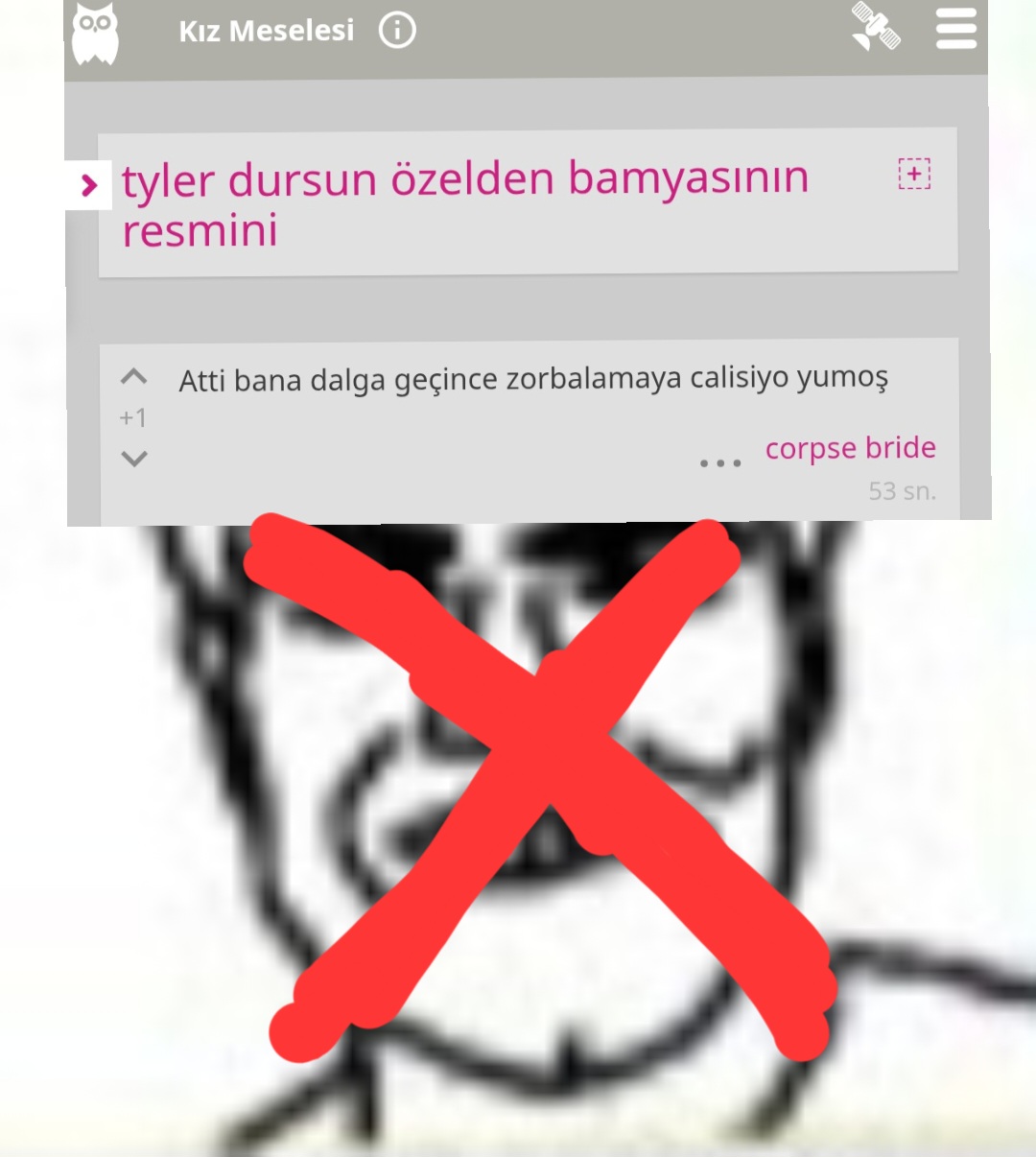


 hay ızdırabını
 pmden yavşadığın karıdan red yiyince
 dümdüz karı fotosu
 rang ve kurtcocain kardeslerimin çaylağı
 kurcoconun çaylağı kalkmazsa
 biz burda sözlük reyisçiliği oynamıyoruz
 sözlük karısının entrysine yanıt vermem
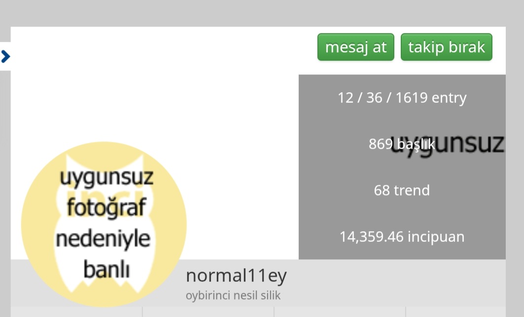
 ayın elemanı seçilmiş
 normal11ey kardeşim seni unutursak
 cccrammsteinccc seni esefle kınıyorum
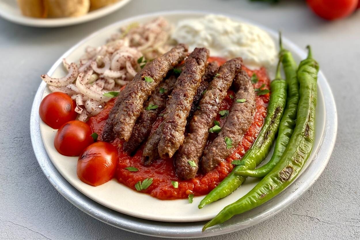
 herkes şu anda yemek istediğini yazıyor
 şadiye amansızoğlu